Products & Services

Recombinant proteins

FGF family and Receptors
Potential Therapeutic Target

Antibiotic Resistance Relative Proteins

FGF family and Receptors

Fibroblast growth factors (FGFs) regulate various developmental and metabolic processes, including cell proliferation, differentiation, angiogenesis, wound healing, nerve regeneration, chronic inflammation, and cancer growth. The FGF family consists of 22 members that share the β-trefoil fold despite their relatively low sequence identities (13–71%). The β-trefoil fold contains 12 β-strands that form 6 two-stranded β-hairpins (i.e., β1-β12, β2-β3, β4-β5, β6-β7, β8-β9, and β10-β11). Hairpins are arranged in a pseudo three-fold symmetry: the first, third, and fifth β-hairpins form a barrel that is covered by a triangular cap consisting of the second, fourth, and sixth β-hairpins.

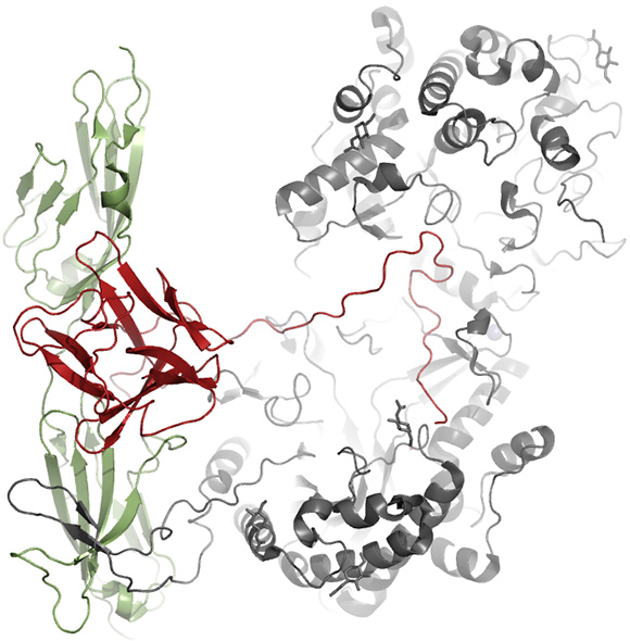
FGFs are divided into FGF1, FGF4, FGF7, FGF8, FGF9, FGF11, and FGF19 subfamilies depending on their sequence similarities. Except for the intracellular FGF11 subfamily, FGFs form complexes with FGF receptors (FGFRs) on the cell membrane, which are generally composed of three immunoglobulin (Ig)-like extracellular domains, a transmembrane domain, and a cytoplasmic tyrosine kinase domain. FGFs bind to the interface between the second and third Ig-like ectodomains to induce receptor dimerization, leading to the phosphorylation of tyrosine residues in the cytoplasmic domain to stimulate signaling pathways.

The canonical FGFs (FGF1, FGF4, FGF7, FGF8, and FGF9 subfamilies) have a positively charged sector clustered by lysine and arginine residues and thus display avidity for negatively charged heparin/heparan sulfate proteoglycans (HSPGs) present on the cell surface or in the extracellular matrix. Therefore, canonical FGFs form ternary complexes with HSPG and FGFR, and act in the vicinity of cells as paracrine and/or autocrine factors. Conversely, FGF19 subfamily members (FGF19, FGF21, and FGF23) have no apparent HSPG-binding site on their surfaces and thus perform their physiological roles in an endocrine manner. They are released from the extracellular matrix and reach remote target organs through the bloodstream.

Complex model structure of
FGFR1c/FGF21/KLB

Cat. # Molecule Species Tag Expression

Host

Product description Quality validation
TODD-B-001 FGF21 human None E. coli hFGF21 Protein, w/o tag SDS-PAGE, BLI
TODD-B-002 FGF21 human None E. coli Endotoxin free hFGF21 Protein, w/o tag SDS-PAGE, BLI

Potential Therapeutic Target
DJ-1 implicated in the pathogenesis of parkinson’s disease

DJ-1, which is extensively expressed in the testis and moderately in other tissues, was first identified as a novel candidate of the oncogene product that transformed mouse NIH3T3 cells in cooperation with activated ras. After the first identification, various physiological roles of DJ-1 have been unveiled. It has been shown that DJ-1 is a circulation tumor antigen in breast cancer, in which DJ-1 is secreted from cells to serum. DJ-1 was also characterized as a protein that regulates an RNA-protein interaction and positively modulates the androgen receptor (AR). Remarkably, the loss of function mutations in DJ-1 gene was revealed to be responsible for the autosomal recessive early-onset Parkinson’s disease (PD). Compatible with diverse cellular functions, human DJ-1 has at least three distinct biochemical activities: chaperon activity, protease activity, and glyoxalase activity.

Cat. # Molecule Species Tag Host Product escription Quality validation
THD001-H DJ-1 human His-tag E. coli Human FGF-21 Protein, His tag SDS-PAGE, Analytical SEC
THD001-O DJ-1 human E. coli Human FGF-21 Protein SDS-PAGE, BLI

Antibiotic resistance related β-lactamases

DJ-1, which is extensively expressed in the testis and moderately in other tissues, was first identified as a novel candidate of the oncogene product that transformed mouse NIH3T3 cells in cooperation with activated ras. After the first identification, various physiological roles of DJ-1 have been unveiled. It has been shown that DJ-1 is a circulation tumor antigen in breast cancer, in which DJ-1 is secreted from cells to serum. DJ-1 was also characterized as a protein that regulates an RNA-protein interaction and positively modulates the androgen receptor (AR). Remarkably, the loss of function mutations in DJ-1 gene was revealed to be responsible for the autosomal recessive early-onset Parkinson’s disease (PD). Compatible with diverse cellular functions, human DJ-1 has at least three distinct biochemical activities: chaperon activity, protease activity, and glyoxalase activity.β-Lactams (penicillins, cephalosporins, and carbapenems) are the most widely used antibiotics to treat bacterial infections. Clinical application of β-lactams has been accompanied by the emergence of bacterial resistance to these antibiotics, which is a great threat to public health. Expression of β-lactamases is a prevalent resistance mechanism of bacteria to β-lactams. β-Lactamases inactivate β-lactams by hydrolyzing the amide bond in the β-lactam ring, the core structure of β-lactams. They are grouped into four classes, A, B, C and D, on the basis of sequence homology among which class A, B, and D are serine β-lactamases and metallo-β-lactamases belong to class B. Although class C β-lactamases along with class A enzymes are the most commonly encountered of the four classes in clinics, class C β-lactamases are more problematic than class A enzymes. Class C β-lactamases can confer resistance to cephamycins (cefoxitin and cefotetan), penicillins, and cephalosporins and are not significantly inhibited by clinically used β-lactamase inhibitors. In contrast, class A β-lactamases are not able to confer resistance to cephamycins and the enzymes are generally susceptible to inhibition by clinically-used inhibitors.

The development of inhibitors against β-lactamases is an effective strategy to cope with the β-lactamase-mediated antibiotic resistance since β-lactams maintain their antibiotic activity in the presence of inhibitors that block β-lactamases. In fact, three classical β-lactam inhibitors (clavulanate, sulbactam, and tazobactam) sharing the β-lactam backbone are clinically used in combination with β-lactam antibiotics (e.g. amoxicillin-clavulanate, ticarcillin-clavulanate, ampicillin-sulbactam , piperacillin-tazobactam, and cefoperazone-sulbactam). These clinical inhibitors are especially active on class A enzymes, displaying much less or no effect on other β-lactamaases. Furthermore, β-lactamases resistant to the β-lactam inhibitors are emerging, which highlights the need to develop non-β-lactam inhibitors with broad-spectrum efficacy.

Class C β-lactamase

& Antibiotics candidates

Cat. # Molecule Species Tag Host Product escription Quality validation
THD001-H DJ-1 human His-tag E. coli Human FGF-21 Protein, His tag SDS-PAGE, Analytical SEC
THD001-O DJ-1 human E. coli Human FGF-21 Protein SDS-PAGE, BLI